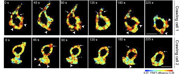
To measure localistion of RhoA activity, the activity in the pixels in the leading edge of the cells was compared to the activity of the pixels in all other edges of the cell. The binomial test was used to measure significance, since in the null hypothesis, the leading edge is equally likely to have either more or less activity compared to the rest of the cell. Th centrod of the cell was taken to be its centre and the movement of the centroid from frame-to-frame was used to calculate the motion. The position of the boundary relative to the centroid was also found as a function of the angular position around the cell. This parameterization allowed the motion of the cell boundary relative to the centre of the cell and the activation to be found as a function of angular position around the cell. The angular position was computed relative to the direction of motion, so (for example), the change in boundary shape and activation of the front of the cell can be readily found.
Updated April 2nd 2020, 05:05